
Chicken autopsy technique
2022-04-02
Qingdao Zhongren Animal Medicine Co., Ltd
Zhongren kinetic drug
The treatment of chicken disease focuses on early treatment and quasi-treatment. In production, due to the small number of initial diseases and the small number of dead chickens, some chicken farmers often dispose of the dead chickens at will, resulting in delays in early treatment and losses. Some of them are far away from the veterinary station or delayed due to the fact that the dead chicken carcasses are sent to the clinic for decomposition, which makes the diagnosis inaccurate and delays the treatment, resulting in losses. Therefore, it is necessary for chicken farmers to master certain techniques for autopsy of chicken carcasses. Once a disease or dead chicken is found and cannot be diagnosed and treated in time, it can be autopsy by itself, and detailed autopsy records can be made, and then the autopsy records can be brought to the veterinarian for diagnosis and treatment.
Dead chickens should check the external changes of the body before dissecting the body cavity. Focus on the body's fat and thin, the color of the face, crown, and beard, whether there are secretions and secretions in the eyes, nose, and mouth, and whether there are tumors and ectoparasites.
Dissection of 2. Body Cavity
First, soak the body with water to prevent feathers flying and sticky hands from affecting the operation, then cut the skin between the thigh and the abdominal wall, and press the thigh to both sides until the femoral head is exposed, so that the body is balanced and supine. Cut the abdominal wall skin across all openings, lift and peel the skin to the head side, and pay attention to the subcutaneous and muscle for bleeding, necrosis and discoloration. At the end of the back abdomen, cut the ribs forward from both sides along the incision, and then lift the sternum with force. At this time, the thoracic and abdominal organs can be exposed. Pay attention to the presence of water, exudate or blood, and observe the location of each organ for abnormalities.
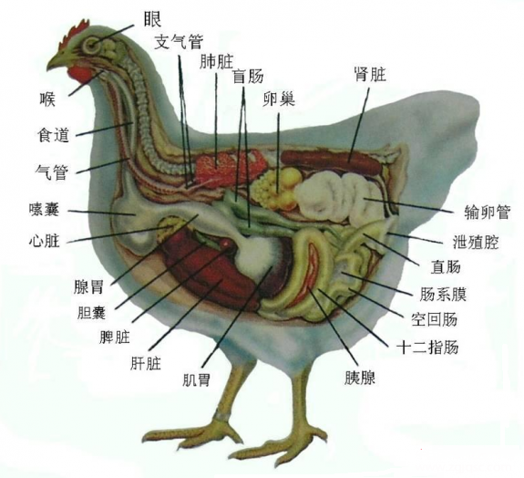
3. organ examination
1. Visceral examination (1) Cut the esophagus between the glandular stomach and the esophagus, and then take out the glandular stomach, muscular stomach, intestinal canal, liver, spleen and pancreas in sequence.
(2) Cut the glandular stomach, pay attention to the presence of parasites, the number, color and state of glandular gastric mucosal secretions; glandular gastric papilla, around the papilla, glandular stomach and esophagus, glandular stomach and muscle stomach junction with bleeding, ulceration. Then cut off the muscle stomach, peel off the cuticle film and pay attention to the presence of parasites. Then cut the intestine longitudinally. Check the contents and mucosal status, whether there are parasites and bleeding, ulceration, intestinal wall for tumors, nodules. Note whether the two blind tubes at the back end of the intestine are swollen and the hardness of the cecum, the state of the mucosa and the nature of the contents. Note whether the cloaca changes.
(3) The spleen is located on the left medial side of the muscle stomach and is round. Pay attention to its color, size, hardness, and whether there is bleeding.
(4) The liver is divided into left and right leaves. Pay attention to the color, size and texture of the liver, whether there is tumor, hemorrhage and necrotic foci. Pay attention to the size and color of the gallbladder.
(5) The kidney is attached to the renal fossa on both sides of the lumbar spine. It is brittle and difficult to be taken out. It can be checked in situ. Focus on the examination of kidney volume, color, whether there is bleeding, necrosis, cut surface whether there is blood outflow, whether there is white uric acid salt deposition. The ovary is located on the left side, pay attention to its size and follicular status. Fallopian tubes can be dissected in situ.
(6) The heart and lungs can be examined in situ. The heart focuses on checking the presence of bleeding points in the crown, endocardium and epicardium, the amount and state of pericardial contents, the presence of blood accumulation in the heart cavity, the color and viscosity of blood accumulation. Lung attention to check the size of the lung, color, whether there is necrosis, nodules and cut surface status.
2, neck organ examination
Turn the head of the chicken towards the autopsy, cut the beak angle to open the mouth, cut the tongue, esophagus and crop, and pay attention to the color, state, smell and mucosal properties of the contents of the crop. Then cut open the larynx, trachea, bronchus, pay attention to the trachea with or without exudate and the amount of exudate, color, state and so on.
3, peripheral nerve, focus on sciatic nerve
At the back of both thighs, the muscles are peeled off to separate the sciatic nerve in white bands or lines. Unilateral sciatic nerve enlargement often occurs in chickens with neurological Marek's disease.
Description and record of 4. autopsy results
1. Measure the length, width and thickness of the diseased organ with a ruler, in centimeters.
2. Use the real object to describe the size and shape of the lesion, but do not use too much disparity, and use the local familiar objects. For example, when indicating a circular volume, large millet grains, large peas, large walnuts, etc. When indicating an ellipse, large soybeans and pigeon eggs can be used. When indicating the area, one cent and five cent coins can be used. When indicating the shape, round, oval, linear, strip, dot, spot, etc.
3. When describing the color of the lesion, if it is a mixed color, the secondary color should be in the front and the main color should be in the back, such as bright red, purple red, gray white, etc. The color can also be described in kind, such as bluestone, red wine, marble and mottled.
4. When describing hardness, it is often described as hard, solid, fragile and soft, and it can also be described as loose and dense.
5. When describing elasticity, rubber samples, dough samples and jelly samples are commonly used. In addition, the autopsy records should also indicate the breed, age, feed, vaccine usage and symptoms before death of sick chickens.
After the autopsy is completed, pay attention to the body, feathers, blood and other objects buried or burned. Disinfect the exposed skin of the autopsy tools and autopsy personnel with disinfectant, and change the clothes and shoes of the autopsy personnel to prevent the spread of pathogens.
Key words:
Contact Us
Administrative service telephone:0532-85212368
Sales Advisory Phone:0086-532-85212381
E-mail:zhongrenyaoye@126.com
Address: No.6 Road, Jiaoxi Industrial Park, Jiaozhou City, Qingdao, Shandong Province

Qingdao Zhongren Animal Medicine Co., Ltd
Zhongren Aquatic Products